A basal cell carcinoma (BCC) is a type of slow-growing skin cancer. It is a malignant tumour which rarely metastasises, but can be locally destructive.
Risk Factors
- UV light exposure is thought to be the major aetiological cause, particularly exposure in childhood. Tanning/using sun beds
- Fair skin/blue eyes/blond hair
- Immunosuppression
- Inherited syndromes
- Gorlin syndrome is a rare condition which increases the risk of basal cell cancer, as well as other tumours such as medulloblastomas.
Clinical Features
- Can be shiny or pearly
- Presents usually as plaque or nodule
- May also present as non-healing wounds or scabs
- Visible telangiectasia on the surface
- Do not produce keratin so they can appear quite translucent. They can, however, be pigmented, especially in patients with darker skin
- Umbilicated centre
- Tend to occur in sun exposed areas e.g. face, ears, neck, chest etc.
- If left untreated, the BCC can ulcerate and increase in size. In this scenario, they typically form a rolled, pearly edge with a crater in the middle.
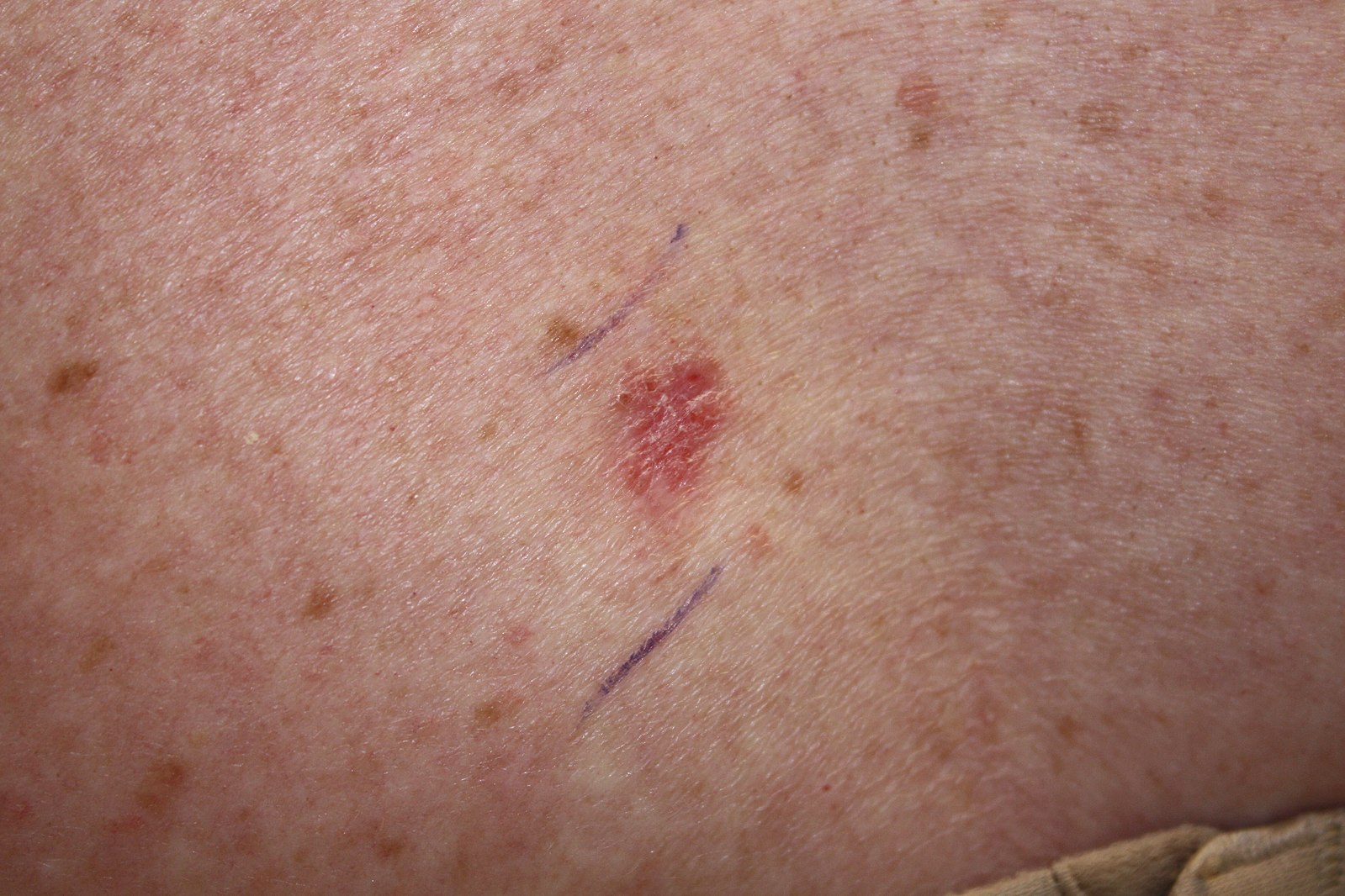
Kelly Nelson, M.D., (Photographer), CC0, via Wikimedia Commons
Superficial Basal Cell Carcinoma
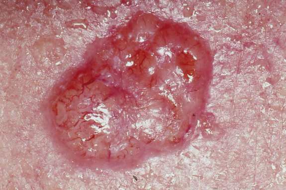
No machine-readable author provided. John Hendrix assumed (based on copyright claims)., Public domain, via Wikimedia Commons
Nodular Basal Cell Carcinoma
There are many different sub-types of BCC:
- Nodular: The most common type of facial BCC, whereby individuals have a pearly, smooth nodule.
- Superficial: These present as plaques and may have scales
- Morephoeic: These are usually in the mid-facial area and tend to have a waxy surface. They are able to infiltrate cutaneous nerves (perineural invasion).
- Basosquamous: These are a mix of BCC and squamous cell carcinomas and tend to be more aggressive.
Investigations
The mainstay of investigation is to biopsy the lesion and histologically confirm presence of BCC.
Management
- Excision Biopsy: Surgical excision is the recommended treatment with usually 3-5mm tumour margin, although this margin can increase for larger BCCs.
- Mohs Micrographic Surgery: This is where a lesion is excised layer-by-layer, and each layer is examined microscopically. Excision continues until tumour is no longer visible on microscopy. This can be useful for sites where you are trying to conserve as much tissue as possible e.g. facial lesions.
- Topical creams
- Imiquimoid: This is an immunomodulator that can be used for superficial BCCs
- Fluorouracil cream (5-FU): This is a cytotoxic drug which again, can be used for superficial lesions
- In patients that do have advanced or metastatic BCC, Hedgehog pathway inhibitors such as vismodegib can be used.
- Radiotherapy: Patients who are unsuitable for excision/Mohs may be offered radiotherapy.
References
https://dermnetnz.org/topics/basal-cell-carcinoma/
https://www.uhb.nhs.uk/Downloads/pdf/CancerPbBasalCellCarcinoma.pdf
https://www.cancerresearchuk.org/about-cancer/other-conditions/gorlin-syndrome
https://www.skincancer.org/skin-cancer-information/basal-cell-carcinoma/
https://gpcpd.heiw.wales/clinical/skin-cancer/basal-cell-carcinoma/
https://onlinelibrary.wiley.com/doi/full/10.1111/bjd.20524