Melanomas are a type of skin cancer which derive from melanocytes, the pigment producing cells found in the epidermis of the skin.
Risk Factors
- UV light
- Family history
- Having many moles
- Fair skin, although melanoma can still develop in darker skin tones
- History of sunburn
- Immunosuppression
Types
- Superficial spreading melanoma
- The most common type of melanoma
- Usually flat
- Slow growing and progressively changing lesion
- Nodular melanoma
- Raised above the skin and tend to be friable
- Usually blue-black in colour
- Lentigo maligna melanoma
- Tends to present as a macule (flat, discoloured lesions)
- Usually found on skin which is chronically exposed to sunlight e.g. head and neck
- Acral lentiginous melanoma
- Form of melanoma which appears on the palms and soles
- Subungual Melanoma
- Present on the nail unit – can appear as a dark pigmented line
- Spitzoid melanoma
- A Spitz naevus is a benign skin lesion
- A Spitzoid melanoma is a malignant melanoma which shares histological similarities to the Spitz naevus.
It’s important to remember that melanoma can develop anywhere on the body, including mucous membranes and in the retina. Additionally, melanoma has the potential for metastasis. It does this by invading through the epidermis, down to the dermis, and then ultimately being able to spread via lymphatics/blood vessels.
Clinical Features
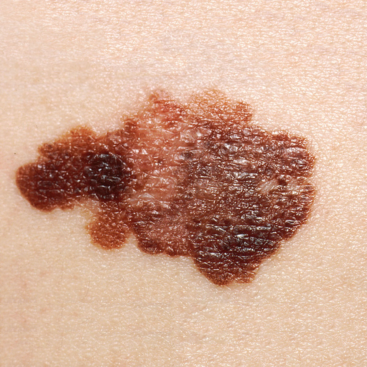
The ABCDE rule is a simple way of knowing how to identify a melanoma. If the following features are present in a skin lesion, there should be a high clinical suspicion for melanoma:
- Asymmetrical shape: Can you draw a line through the middle and it’s the same on both sides of the line?
- Border: Is there an irregular border?
- Colour: Are there irregular colours in the lesion? Melanoma can have various colours e.g. black, dark brown, blue, tan etc. They may also have no colour at all (amelanotic melanoma)
- Diameter: Is it greater than 6mm?
- Evolution: Has the lesion changed in size or shape since it first appeared?
Unknown photographer, Public domain, via Wikimedia Commons
Melanoma
A mole which is also itching or bleeding, or a new pigmented line under the nail should warrant urgent referral as well.
We worry about a melanoma as it has potential for metastasis.
Differential Diagnosis
- Pigmented basal cell carcinoma
- Seborrheic keratosis: Waxy, scaly, and slightly raised lesions which and are totally benign
- Benign melanocytic naevus (mole)
Investigations
UK guidelines state that referred pigmented skin lesions should be assessed using dermoscopy.
Bloods
- Vitamin D: Low vitamin D is associated with higher Breslow thickness. Additionally, some literature has linked vitamin D as being anti-proliferative for some cases of melanoma.
- LDH: Elevated in metastasis
- ALP: Elevated in instances of bone metastases
- Liver function test: Liver metastases may result in derangement
Imaging
- CT scan head, thorax, abdomen and pelvis: Whole-body and brain CT with contrast can be offered based on the staging of the melanoma
- Whole body and brain MRI may be used instead of CT in certain cases e.g. children/young adults or pregnant women
Special Tests
- Sentinel lymph node biopsy may also be needed depending on the stage of the disease/features of the disease e.g. Breslow thickness >1mm.
Staging
Melanoma is staged using Clark’s level and Breslow’s thickness.
- Breslow’s Thickness: This is a measurement of the depth or thickness of the tumour in mm, and is done by a pathologist. It correlates to how aggressive the tumour will be, and hence the prognosis. The thicker a melanoma, the higher the likelihood of metastasis.
- Clark’s Level: Used in conjunction with Breslow’s thickness for staging, and again, the higher the level the higher the likelihood of metastasis. The scale has 5 levels:
- Level 1: Melanoma cells are only in the epidermis. This is also known as melanoma in situ.
- Level 2: Melanoma cells are in the papillary (superficial) dermis
- Level 3: Melanoma cells extend to the junction between the papillary and reticular dermis
- Level 4: Melanoma cells are in the reticular (deep) dermis
- Level 5: Melanoma cells are in the subcutaneous tissue
- TNM Staging: This is a common staging system for many cancers and stands for Tumour, Node, Metastasis.
- Tumour: This is the thickness of the melanoma (mm). Some of these stages are further split up into a and b, but for sake of simplicity we’ve only included the overarching number.
- Tis: Melanoma in situ
- T0: No melanoma
- T1: <1mm
- T2: 1mm-2mm
- T3: 2mm-4mm
- T4: >4mm
- Node: Metastases to lymph nodes
- N0: No melanoma in local lymph nodes
- N1: Melanoma in one lymph node, or in-transit/microsatellite/satellite
- N2: Melanoma in 2-3 lymph nodes or in 1 lymph node + in-transit/microsatellite/satellite
- N3: Melanoma in 4> lymph nodes or in 2/3 lymph node + in-transit/microsatellite/satellite
- Metastasis
- M0: No spread
- M1: Metastasis present in another part of the body
These 3 elements of staging then combine to give a stage from 0 to IV. It’s hard to remember all of this for your exams but if you remember Stage 0-stage II confers localised disease i.e. N0, M0. Stage III involves the nodes, and stage IV involves nodes and any distant metastases.
Lymph Nodes Metastasis
- Microsatellite: Melanoma cells visible within 0.3mm from primary melanoma site
- Satellite: Melanoma visibly spread <2cm from primary melanoma site
- In-transit: Melanoma within lymphatic vessels > 2cm from primary melanoma site
Management
Lesions which cause high clinical suspicion for melanoma should be referred on the urgent 2-week wait referral pathway in the UK. All lesions suspected to be melanomas are excised – this is then biopsied for staging via Breslow’s thickness and Clark’s levels.
Excision
Excision is usually the management option of choice for localised disease, with a margin of at least:
- 0.5cm for stage 0
- 1cm for stage I
- 2cm for stage II
For more advanced melanoma e.g. IIIB -> IIID (3B -> 3D), therapeutic lymph node dissection are sometimes offered alongside excision, but it is not routinely offered unless there are presence of factors that would make it difficult to treat recurrent lymph node disease e.g. head and neck melanoma or where regular follow-up would not be possible.
Imiquimoid
Imiquimoid is an immune response modifier and can be used as a treatment option is only really considered for stage 0 melanoma, and only in instances where surgery is unacceptable e.g. it would cause morbidity/disfigurement. It can also be used in more advanced cases e.g. superficial skin metastases, but this is usually for palliative purposes and is not a curative option.
Systemic Treatments
- Immunotherapy
- Nivolumab + Ipilimumab are a common combination of immunotherapy that can be used to treat patients with unresectable stage III or untreated stage IV melanoma.
- Alternatives include pembrolizumab or nivolumab single agent therapy. Immunotherapy carries a risk of immunotherapy induced toxicities.
- BRAF/MEK Inhibitors
- BRAF inhibitors are a type of targeted cancer treatment that target cells with BRAF mutations.
- Some melanomas have mutated BRAF genes (and thus BRAF proteins) that promote their growth, and so this can be targeted as part of cancer therapy.
- There is also a MEK gene which works with the BRAF gene, so the MEK gene can also be treated with MEK inhibitors to help shrink tumours/slow their growth.
- BRAF analysis of melanoma tissue is carried out of for individuals with stage IIC -> IV melanoma.
- NICE suggest in instances where immunotherapy cannot be used, encorafenib (a BRAF inhibitor) can be used in conjunction with binimetinib (a MEK inhibitor).
Complications
Metastasis is a serious concern in malignant melanoma. Common sites of metastasis include the
Prognosis
- Stage I: Close to 100% 5-year survival rate
- Stage II: 80% 5-year survival rate
- Stage III: 70% 5-year survival rate
- Stage IV: 30% 5-year survival rate
It’s important to bear in mind the statistics for stage IV do not take into consideration the age of the patients diagnosed.
References
https://training.seer.cancer.gov/melanoma/abstract-code-stage/staging.html
https://www.ncbi.nlm.nih.gov/books/NBK482163/
https://dermnetnz.org/topics/spitz-naevus/
https://dermnetnz.org/topics/spitzoid-melanoma/
https://www.bad.org.uk/library-media%5Cdocuments%5CMelanoma_2010.pdf
https://www.ncbi.nlm.nih.gov/pmc/articles/PMC3756353/
https://www.nice.org.uk/guidance/NG14/chapter/1-Recommendations#assessing-melanoma-2
https://www.cancerresearchuk.org/about-cancer/melanoma/stages-types/tnm-staging
https://www.nice.org.uk/guidance/ng14/chapter/Recommendations#managing-vitamin-d-levels-and-concurrent-drug-treatment
https://www.cancer.org/cancer/melanoma-skin-cancer/treating/targeted-therapy.html
https://www.ncbi.nlm.nih.gov/pmc/articles/PMC3897580/
https://pubmed.ncbi.nlm.nih.gov/26016970/
https://www.cancerresearchuk.org/about-cancer/melanoma/survival