Inflammation of the pancreas gland can occur for a number of reasons, resulting in a set of clinical features and signs.
Causes
There are two main causes of acute pancreatitis: gall stones and alcohol. Other causes do exist which are best remembered by the mnemonic “I GET SMASHED”:
I: Idiopathic
G: Gallstones
E: Ethanol
T: Trauma
S: Steroids
M: Mumps
A: Autoimmune
S: Scorpion sting
H: Hyperlipidaemia/hyperparathyroidism/hypercalcaemia
E: ERCP
D: Drugs (E.g. tetracyclines, furosemide)
Pathophysiology
The exact mechanism by which pancreatitis is initiated is unclear. However, in simple terms it is thought an initial precipitating event will lead to activation of zymogens and subsequent autodigestion.
With regards to gallstone induced pancreatitis, it is thought that gallstones can lead to an obstruction of the biliary-pancreatic duct. This increases the pressure in the pancreatic duct which causes activation of pancreatic zymogens (inactive proteases) such as trypsinogen into trypsin. Trypsin is capable of further activating trypsinogen. Such high levels of active proteases can lead to pancreatic autodigestion and subsequent pain, inflammation and oedema.[i]
Clinical Features
- Upper/central abdominal pain which radiates to the back (the pancreas is a retroperitoneal structure and the retroperitoneum can become involved).
- Pain may be relieved when sitting forward and is worse when lying on the back.
- Nausea/vomiting
- Systemic upset
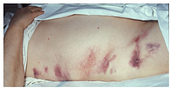
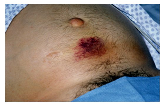
There may also be bruising if there has been autodigestion of nearby blood vessels. This presents as periumbilical bruising (Cullen’s sign) or flank bruising (Grey Turner’s sign) and is a sign of necrotising pancreatitis.
Herbert L. Fred, MD and Hendrik A. van Dijk, CC BY 2.0 , via Wikimedia Commons
Grey Turner's Sign
Cullen's Sign
Investigations
Bedside
- History and Examination: History of excessive alcohol use/gallstone history?
- Fluid Status Examination
- ECG
Bloods
- FBC, U&E, TFT, LFT, Clotting: Baseline
- Bone Profile: For calcium as it can cause pancreatitis
- CRP: Inflammatory marker
- Amylase: If this is three times the upper limit, it is highly suggestive of pancreatitis. However, it is important to note that amylase may be normal despite pancreatitis. Additionally, serum amylase may be elevated in other conditions as well such as gastrointestinal perforation.
- Lipase: This is more sensitive for pancreatitis
- ABG: PaO2 is part of risk assessment and is important to monitor to check for hypoxia
Imaging
- CXR: An erect chest x-ray is important, particularly if amylase is raised as this can be due to a gastrointestinal perforation.
- Abdominal ultrasound: ?Gallstones
- CT with contrast: The main imaging technique to visualise the pancreas and assess if there is any pancreatic necrosis or evidence of complications such as a pseudocyst or abscess.
- MRCP: ?Gallstones
Risk Assessment
Several risk assessment criteria exist for acute pancreatitis including the Acute Physiology and Chronic Health Examination (APACHE)-II and Glasgow criteria.
The Glasgow criteria helps to assess the severity of pancreatitis and whether or not a patient may require HDU/ICU transfer. The Glasgow Criteria can be remembered using the mnemonic PANCREAS, and are as follows:
- PaO2: <8
- Age: >55
- Neutrophilia: WBC>15
- Calcium: <2
- Renal: Urea >16
- Enzymes: LDH >600; AST >200
- Albumin: <32
- Sugar: Glucose >10
A score of 3 or more within 48 hours of symptom onset suggests severe pancreatitis.[ii]
Management
Supportive
- Hydration: Fluid losses occur in pancreatitis through vomiting and oedema – it is crucial to replenish fluid losses. IV fluids are usually administered (crystalloids).
- Monitor Fluids: Urinary catherisation and subsequent monitoring of urinary output.
- Nutrition: Patients should only be made nil-by-mouth if there is a reason e.g. severe vomiting – patients with mild pancreatitis may be able to have oral nutrition. Otherwise, enteral feeding is preferred e.g. through a nasogastric tube. Parenteral nutrition is used if patients cannot tolerate enteral feeding.i
- Oral Feeding: Via mouth
- Enteral Feeding: Feeding via the gastrointestinal tract but without using the mouth e.g. a nasogastric feeding tube, gastric feeding tube, nasojejunal feeding tube etc.
- Parenteral Feeding: Feeding intravenously
- Analgesia: Opiates are the drug of choice such as tramadol. Morphine and diamorphine may be avoided due to risk of these drugs causing spasm of the sphincter of Oddi and subsequent increased pressures within the pancreatic ductal system though evidence for this is limited.
- Anti-emetics
- Antibiotics: If concerns about infection
Definitive
Endoscopic retrograde cholangiopancreatography (ERCP): This can be performed to remove gallstones. An endoscope is passed from the mouth into the stomach and then into the duodenum. The scope can be passed through the ampulla of Vater into the pancreatic ductal system or the bile duct in order to remove gallstones or stent the pancreatic duct.
Cholecystectomy may also be performed to prevent future episodes.
Seek Help
Consider contacting HDU/ITU
Difference Between ERCP and MRCP
Endoscopic retrograde cholangiopancreatography or ERCP, is a procedure which can be diagnostic and therapeutic. It involves passing an endoscope from the mouth and ultimately into either the pancreatic or bile ducts to dilate the ducts with a stent, remove gallstones etc.
Magnetic resonance cholangiopancreatography or MRCP, is an imaging technique that uses MRI to image the pancreatic and biliary ductal system. In the image below, you can see the gallstones that have become impacted into the common bile duct (labelled b).
Complications
- Hypocalcaemia: Whilst hypercalcaemia can cause pancreatitis, pancreatitis is able to cause hypocalcaemia. Theories have been proposed as to how this happens – one suggests that pancreatitis leads to autodigestion of fat surrounding the pancreas which causes the release of fatty acids which subsequently bind to calcium ions to form calcium salts, resulting in hypocalcaemia.[iii]
- Acute Kidney Injury: Due to dehydration
- Pancreatic pseudocyst: Collection of fluid containing pancreatic enzymes/blood/tissue
- Pancreatic abscess
- Pancreatic necrosis and subsequent infection
- Sepsis
References
[i] Wang G, Gao C, Wei D, Wang C and Ding S. Acute pancreatitis: Etiology and common pathogenesis. [internet]. 2009. [cited 11th August 2019]. Available from: https://www.ncbi.nlm.nih.gov/pmc/articles/PMC2665136/
[ii] Oxford Handbook
[iii] Ahmed A, Azim A, Gurjar M and Baronia AK. Hypocalcemia in acute pancreatitis revisisted. [internet]. 2016. [cited 11th August 2019]. Available from: https://www.ncbi.nlm.nih.gov/pmc/articles/PMC4810896/