Several types of oesophageal cancer exist such as adenocarcinoma or squamous cell carcinoma of the oesophagus.
Types
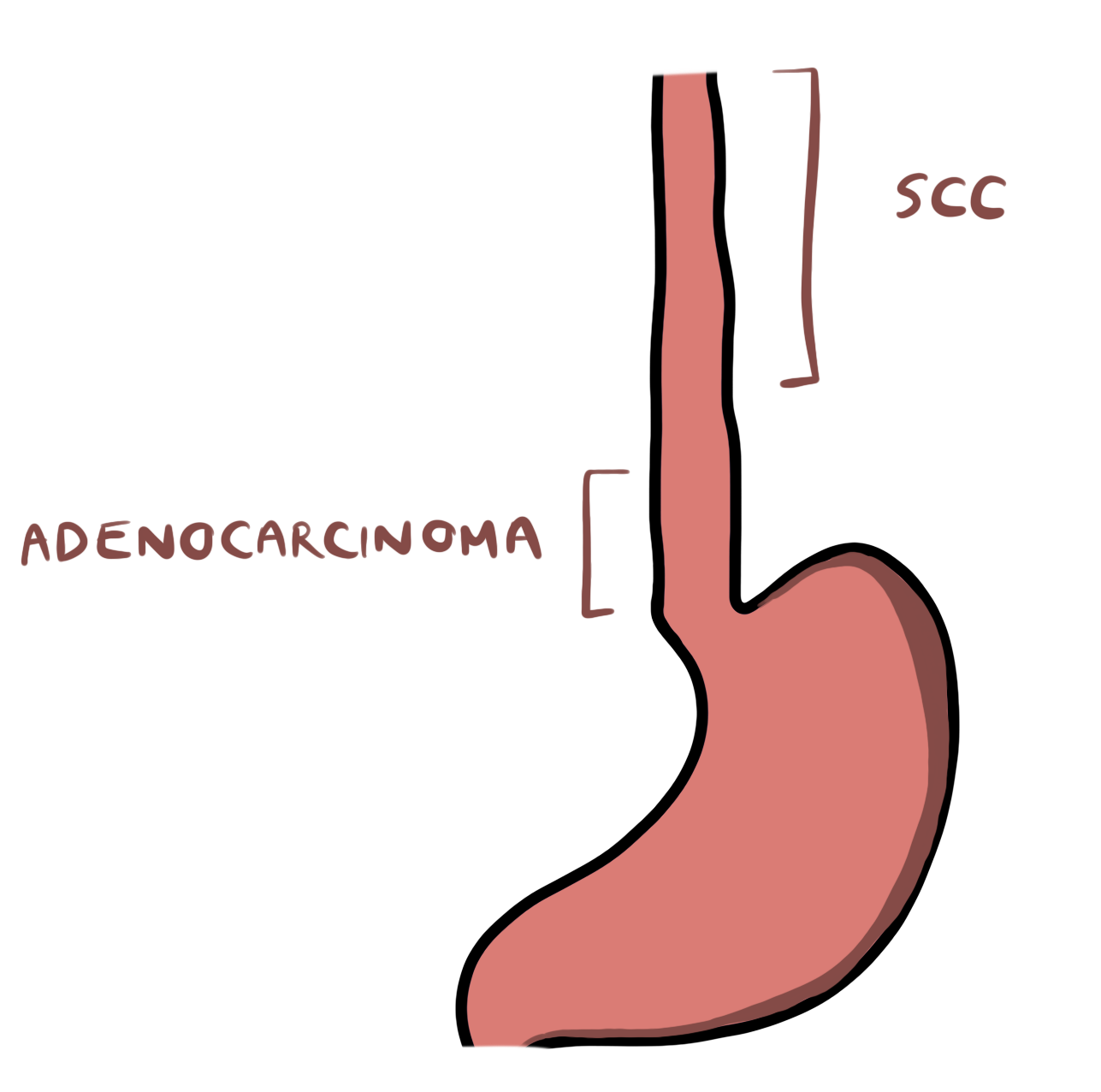
- Adenocarcinoma: Typically develops from the epithelium of the lower third of the oesophagus and is associated with a history Barrett’s oesophagus and gastro-oesophageal reflux disease.
- Squamous cell: Linked to alcohol consumption and tobacco smoking and is usually found in the upper two thirds of the oesophagus.
Copyright Medic in a Minute 2022
Adenocarcinoma and Squamous Cell Carcinoma Common Sites
Epidemiology
Oesophageal is the 8th most common cancer, with the squamous type being more common internationally though the incidence of oesophageal adenocarcinomas is increasing.[i] This rise in adenocarcinomas is likely linked to the rise in obesity, as this is a risk factor for development of adenocarcinoma.
Risk Factors
Adenocarcinoma:
- Barrett’s oesophagus
- Smoking
- Chronic gastro-oesophageal reflux disease
- Obesity
- Zollinger-Ellison syndrome: Rare condition where you have gastrin producing tumours that result in excessive hydrochloric acid secretion
Squamous cell carcinoma:
Clinical Features
- Dysphagia: Usually progressive i.e. starts by affecting solids but will begin to involve liquids very soon after.
- Chest Pain: Food can get stuck which may also cause pain.
- Weight Loss: Due to the dysphagia, and difficulty eating.
- Aspiration: From dysphagia
- Lymphadenopathy
- Fever
- Lethargy
- Hoarse voice: If the tumour infiltrates to the recurrent laryngeal nerve
Endoscopy
The main investigation modality is endoscopy, as it allows visualisation of a tumour. The following outline the NICE criteria for an urgent 2-week wait referral for upper-GI endoscopy.
- Anyone with dysphagia OR
- >55 years + weight loss + one of the following
- Dyspepsia
- Reflux
- Upper Abdominal Pain
Biopsy can be performed to confirm the presence of a carcinoma.[ii]
Staging
Tumour, nodes, metastases (TNM) staging is used to stage oesophageal cancer.
- CT thorax-abdomen and pelvis: Usually the first step to assess the extent of invasion of the tumour as well as lymph node involvement.
- PET Scan: Confirm distant metastases that are suspected on CT.
- Endoscopic ultrasound: To further assess tumour invasion and staging, particularly in advanced tumours. Fine needle aspirations can also be taken of lymph nodes to check for nodal involvement.
- Staging laparoscopy: May also be performed, particularly if a lower oesophageal tumour or a gastro-oesophageal junction tumour in order to assess metastases to the peritoneum or liver.[iii]
Management
Treatment Options
It is good to think of cancer treatment in two major categories – curative intent, or palliative intent.
- Surgery: Provides a chance of cure but the tumour needs to be fairly localised, ideally not infiltrating outside of the oesophageal wall.
- Oesophagectomy
- Endoscopic mucosal resection: For early stage cancers e.g. T1N0
- Chemotherapy
- Neoadjuvant: This is chemotherapy that is used to reduce the tumour load prior to the primary treatment being given. For example, pre-operative chemotherapy.
- Adjuvant: Given after the primary treatment to clear remaining cancer.
- Radiotherapy
- The use of high energy radiation to again, reduce the tumour load.
- Can cause unpleasant side effects when targeting the head and neck region, known as radiotherapy toxicity. Symptoms include
- Mucositis
- Oesophagitis
- Odynophagia
- Loss of taste
- Increased secretions which can cause nausea and vomiting
- Chemoradiotherapy: Combination of chemotherapy and radiotherapy. This can be given alone as the definitive treatment, or before surgery.
- Palliative Therapy: Essentially symptom control only – no curative intent to the treatment. This can involve
- Palliative chemotherapy/radiotherapy
- Oesophageal stents
- Best supportive care: This involves symptom control e.g. managing pain or giving IV fluids where patients have reduced oral intake etc.
Multi-Disciplinary Team
Treatment must always take place under a multi-disciplinary team consisting of physicians, surgeons, clinical nurse specialists, dieticians, speech and language therapists, and more.
Nutritional support is particularly important in these patients, and they make require things such as a gastrostomy.
- Gastrostomy: Tube that goes from the skin, through the stomach wall and into the stomach.
- PEG tubes: Percutaneous Endoscopic Gastrostomy: Inserted endoscopically
- RIG tubes: Radiologically Inserted Percutaneous Gastrostomy: Inserted using X-ray guidance
- Ensures: High calorie milkshake drinks to help reach nutritional and calorific intake
References
[i] https://www.ncbi.nlm.nih.gov/pmc/articles/PMC3769895/
[ii] https://www.nice.org.uk/guidance/ng12/chapter/1-Recommendations-organised-by-site-of-cancer#upper-gastrointestinal-tract-cancers
[iii] https://www.nice.org.uk/guidance/ng83/chapter/Recommendations#assessment-after-diagnosis